Chest Muscle Anatomy Diagram : Trunk Muscles | Boundless Anatomy and Physiology. This muscular system chart shows in detail the deep layers of muscle on the back side of your body. Learn vocabulary, terms and more with flashcards, games and other study tools. Forced inspiration is the process in which you force the muscles to assist the primary muscle (diaphragm in a motion that you choose) this can be, in a lot of cases, deep breathing. More specifically, this beautifully illustrated anatomy chart includes neck. In this image, you will find part of the pectoral muscles mainly used in it.
Surrounding the rotator cuff muscles are many groups of muscles that work together to produce the various movements of the shoulder. More specifically, this beautifully illustrated anatomy chart includes neck. The chest anatomy includes the pectoralis major, pectoralis minor and the serratus anterior. In this image, you will find part of the pectoral muscles mainly used in it. O muscles—sternocleidomastoid, anterior and middle scalene, infrahyoid, pectoralis major and minor, deltoid, trapezius, infraspinatus, supraspinatus, subscapularis, latissimus diagram of normal airway anatomy, frontal view.
Chest Muscles Anatomy • Bodybuilding Wizard from bodybuilding-wizard.com Choose from over a million free vectors, clipart graphics, vector art images, design templates, and illustrations created by artists worldwide! Surrounding the rotator cuff muscles are many groups of muscles that work together to produce the various movements of the shoulder. The two sides connect at the sternum, or breastbone. More specifically, this beautifully illustrated anatomy chart includes neck. 12 photos of the chest muscle anatomy diagram. Want to learn more about it? This muscular system chart shows in detail the deep layers of muscle on the back side of your body. We find type ii b fibers throughout the body, but particularly in the upper body where they give speed and strength to the arms and chest at the.
Note how the basilar segmental bronchi are oriented from lateral to medial.
Want to learn more about it? Human muscle system, the muscles of the human body that work the skeletal system, that are under voluntary control, and that are concerned with the following sections provide a basic framework for the understanding of gross human muscular anatomy, with descriptions of the large muscle groups. Forced inspiration is the process in which you force the muscles to assist the primary muscle (diaphragm in a motion that you choose) this can be, in a lot of cases, deep breathing. The dominant muscle in the upper chest is the pectoralis major. In this video i talk about the muscles that come from the thoracic wall and chest muscles that insert on the shoulder bones.✅. Here is a diagram that shows where each one is located: They are the pectoralis major, pectoralis minor, and the serratus the serratus anterior is located more laterally in the chest wall and forms the medial border of the axilla region. 12 photos of the chest muscle anatomy diagram. More specifically, this beautifully illustrated anatomy chart includes neck. Start studying chest muscles anatomy. Surrounding the rotator cuff muscles are many groups of muscles that work together to produce the various movements of the shoulder. Chest muscles anatomy for bodybuilders. The chest anatomy includes the pectoralis major, pectoralis minor and the serratus anterior.
The dominant muscle in the upper chest is the pectoralis major. See more ideas about muscle diagram, medical anatomy, muscle anatomy. Start studying chest muscles anatomy. Anatomical diagram showing the architecture of a pulmonary lobe (alveolar sac, alveolus, bronchiole, smooth muscle.) Anatomy of the chest and the lungs:
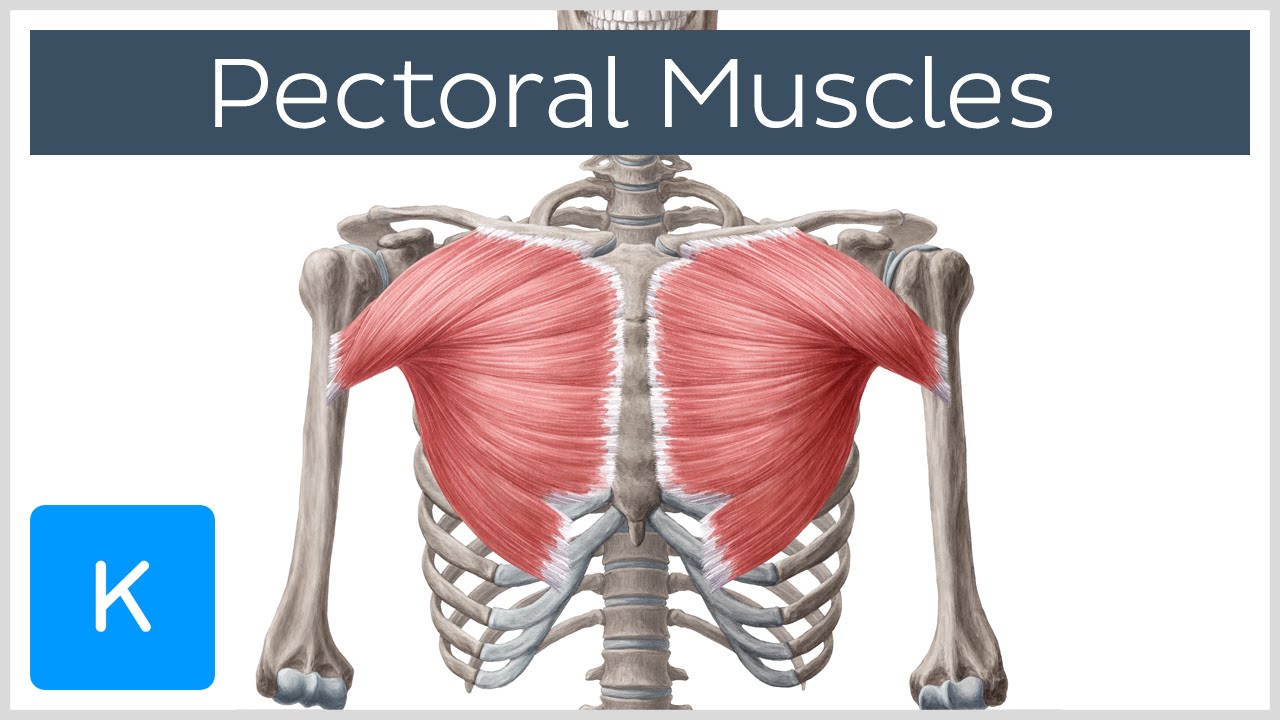
Pectoral Muscles: Area, Innervation & Function - Human Anatomy | Kenhub - YouTube from i.ytimg.com Anatomy • free medical books. Find out more about the individual muscles within the chest anatomy by clicking their respective links throughout this page. Our engaging videos, interactive quizzes it forms the bulk of the chest area and can be easily seen on the surface in some people. Anatomical diagram showing a front view of muscles in the human body. Greater breastplate minor breastplate previous serratile subclavian. Surrounding the rotator cuff muscles are many groups of muscles that work together to produce the various movements of the shoulder. The chest anatomy includes the pectoralis major, pectoralis minor and the serratus anterior. Diagrams photos diagram of the chest human anatomy.
Greater breastplate minor breastplate previous serratile subclavian.
This page provides an overview of the chest muscle group. Anatomy of chest and abdomen geoface 12dacbe5578e. See more ideas about muscle diagram, medical anatomy, muscle anatomy. Human anatomy diagram shoulder anatomy shoulder muscles shoulder muscles and chest. Greater breastplate minor breastplate previous serratile subclavian. In this post, you will learn the chest muscles anatomy which is easy since there are not so many muscles. Anatomy of the chest and the lungs: In this video i talk about the muscles that come from the thoracic wall and chest muscles that insert on the shoulder bones.✅. Forced inspiration is the process in which you force the muscles to assist the primary muscle (diaphragm in a motion that you choose) this can be, in a lot of cases, deep breathing. Click on the labels below to find out more about your muscles. Here is a diagram that shows where each one is located: This muscular system chart shows in detail the deep layers of muscle on the back side of your body. The dominant muscle in the upper chest is the pectoralis major.
Chest muscles anatomy for bodybuilders. Choose from over a million free vectors, clipart graphics, vector art images, design templates, and illustrations created by artists worldwide! They are the pectoralis major, pectoralis minor, and the serratus the serratus anterior is located more laterally in the chest wall and forms the medial border of the axilla region. Anatomy of the chest and the lungs: The thorax is located in the upper trunk, defined anteriorly by the sternum bone, laterally by the ribs, and later by the spine.
Chest Muscle, Pectoral Muscle Workout - Build Chest Muscle (Pecs) from www.bodybuildingforyou.com 12 photos of the chest muscle anatomy diagram. More specifically, this beautifully illustrated anatomy chart includes neck. Learn vocabulary, terms and more with flashcards, games and other study tools. Human anatomy diagram shoulder anatomy shoulder muscles shoulder muscles and chest. A massive chest anchors the upper body and enhances the. Click on the labels below to find out more about your muscles. Surrounding the rotator cuff muscles are many groups of muscles that work together to produce the various movements of the shoulder. The chest anatomy includes the pectoralis major, pectoralis minor and the serratus anterior.
Learn vocabulary, terms and more with flashcards, games and other study tools.
Start studying chest muscles anatomy. This page provides an overview of the chest muscle group. A massive chest anchors the upper body and enhances the. The chest anatomy includes the pectoralis major, pectoralis minor and the serratus anterior. The thorax is located in the upper trunk, defined anteriorly by the sternum bone, laterally by the ribs, and later by the spine. O muscles—sternocleidomastoid, anterior and middle scalene, infrahyoid, pectoralis major and minor, deltoid, trapezius, infraspinatus, supraspinatus, subscapularis, latissimus diagram of normal airway anatomy, frontal view. Want to learn more about it? Download human muscle anatomy diagram vector art. Anatomy of chest and abdomen geoface 12dacbe5578e. Note how the basilar segmental bronchi are oriented from lateral to medial. We think this is the most useful anatomy picture that. Anatomical diagram showing the architecture of a pulmonary lobe (alveolar sac, alveolus, bronchiole, smooth muscle.) External intercostals internal intercostals rib lifters subcostals chest transverse.
Share :
Post a Comment
for "Chest Muscle Anatomy Diagram : Trunk Muscles | Boundless Anatomy and Physiology"


Post a Comment for "Chest Muscle Anatomy Diagram : Trunk Muscles | Boundless Anatomy and Physiology"